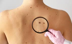
Melanoma is a dangerous form of skin cancer that originates from melanocytes—cells responsible for producing melanin, the pigment that gives color to our skin, hair, and eyes. Although melanoma is less common than other types of skin cancer, it tends to grow and spread more aggressively, making early detection critical.
What Are the Warning Signs of Melanoma?
Melanoma can develop on normal skin or from pre-existing moles. The most common signs are changes in the appearance of a mole or the sudden appearance of a new lesion.
To help recognize early symptoms, dermatologists often refer to the ABCDE rule:
- A – Asymmetry: One half of the mole or lesion does not match the other half.
- B – Border: Edges may be irregular, ragged, notched, or blurred.
- C – Color: The color is not uniform and may include shades of brown, black, pink, red, white, or blue.
- D – Diameter: The mole is larger than 6 mm (about the size of a pencil eraser), although melanomas can be smaller.
- E – Evolving: Any change in size, shape, color, texture, or new symptoms such as itching, bleeding, or crusting.

In addition to these signs, melanoma may appear in unusual places, such as under the fingernails or toenails, on the palms and soles, or even inside the mouth and eyes.
If you notice any of the above signs, or if a mole looks noticeably different from others (“the ugly duckling sign”), seek medical evaluation promptly.
What Causes Melanoma?
Melanoma results from mutations in melanocyte DNA, leading to uncontrolled cell growth. Several factors increase the risk of developing melanoma:
- Ultraviolet (UV) radiation: The leading cause, especially from excessive sun exposure and the use of tanning beds.
- Light skin: Individuals with fair skin, light eyes, and blond or red hair have less melanin and are at higher risk.
- Frequent sunburns: Especially those with blistering sunburns in childhood or adolescence.
- Family history of melanoma: Having a close relative with melanoma increases your risk.
- Personal history of skin cancer: A previous melanoma or other skin cancer raises your chance of recurrence.
- Multiple or atypical moles: Especially dysplastic nevi, which are irregularly shaped or larger than normal moles.
- Weakened immune system: Due to certain medical conditions or immunosuppressive treatments (e.g. after organ transplantation).
- Genetic mutations: Inherited mutations, particularly in the CDKN2A or BRAF gene, may contribute to melanoma.

Melanoma is more common in people over the age of 50, but it can affect younger individuals as well. It occurs more frequently in men than in women, and is particularly prevalent in people living in areas with high levels of UV radiation, such as Australia and New Zealand.
How Is Melanoma Diagnosed?
If a mole or lesion appears suspicious, a dermatologist will carry out a series of evaluations:
- Physical examination: The doctor will assess your skin using a dermatoscope (a magnifying device with a light source).
- Skin biopsy: The definitive method for diagnosing melanoma. A sample of the skin lesion is removed and examined under a microscope to confirm the presence of cancer cells.
- Histopathology: Helps determine key features of the tumor, such as:
– Breslow thickness (tumor depth),
– Presence of ulceration,
– Mitotic rate (rate of cell division).
For patients with suspected advanced melanoma, further imaging tests may be ordered:
- Ultrasound or CT scans: To assess lymph nodes and internal organs.
- PET or MRI scans: To detect distant metastases.
- Sentinel lymph node biopsy: A surgical procedure to determine if the cancer has spread to nearby lymph nodes.
Stages of Melanoma
Staging is crucial for determining the prognosis and treatment plan. Melanoma is classified from Stage 0 (in situ) to Stage IV based on factors like tumor thickness, ulceration, lymph node involvement, and metastasis.
- Stage 0 (in situ): Cancer is confined to the top layer of skin (epidermis). Highly curable with surgery.
- Stage I–II: The tumor is thicker and may be ulcerated but hasn’t spread to lymph nodes. Risk of metastasis increases with tumor thickness.
- Stage III: The cancer has spread to nearby lymph nodes or nearby skin (satellite or in-transit metastasis).
- Stage IV: Melanoma has spread to distant organs such as the lungs, liver, brain, bones, or distant skin. This stage requires more complex, systemic treatments.
The staging system commonly used is the AJCC TNM system, which evaluates:
- T (Tumor): Thickness and ulceration.
- N (Nodes): Involvement of lymph nodes.
- M (Metastasis): Presence of distant spread.
Prognosis of Melanoma
The prognosis of melanoma depends on how early it is diagnosed and treated:
- Stage 0 and I: Excellent prognosis. 5-year survival rates are over 90–99%.
- Stage II: Still treatable with surgery, but survival rates begin to decline (around 70–85% depending on ulceration and thickness).
- Stage III: Moderate prognosis; 5-year survival rates range from 40–78% depending on the extent of lymph node involvement.
- Stage IV: Poorer prognosis due to metastasis, but recent advances in treatment have significantly improved outcomes. Some patients now achieve long-term remission.

Prevention and Early Detection
Although melanoma can be aggressive, it is highly preventable and treatable when caught early. You can reduce your risk by:
- Avoiding excessive sun exposure, especially between 10 a.m. and 4 p.m.
- Wearing protective clothing, hats, and sunglasses.
- Using broad-spectrum sunscreen with SPF 30 or higher, and reapplying every two hours.
- Avoiding tanning beds.
- Regular self-examination: Check your skin monthly using a mirror, and pay attention to new or changing moles.
- Routine skin checks with a dermatologist, especially if you have risk factors.

Melanoma is not just “skin deep.” While it can be a silent and fast-moving disease, awareness and vigilance can make all the difference. Protect your skin, know the signs, and don’t hesitate to consult a doctor if something doesn’t look right.

